Finally, the Findings…
This has taken me far longer to write than I care to admit. What you’re about to read is the facts behind what was occurring in Jersey’s body in the five short years of his life. Dr. Sharon May-Davis and the Equus-Soma team did an incredible job with his disassembly (scientific term) and to honor that you will find a list of just the facts first- without any speculation as to what caused them. I’ve done my best to write in a way that is accessible to most, even if you don’t know every muscle or scientific term. I wanted it to be as digestible as possible but feel free to ask questions!
I put a not-so-little note towards the end of this document with my personal speculations based on the findings and discussions with the team. The only real truth we have is what his body taught us during his disassembly and what we know of his symptoms and behavior during the two years that I owned him.
Here’s the facts–
The Organs:
His adrenal glands were enlarged- much larger than what should be present in a horse of his age. The adrenals were riddled with adrenal adenomas (small benign orange tumors). One of the adrenals that was dissected appeared pale internally and had over twenty adenomas present.
There was superficial plaque on the kidneys. Upon entering the body cavity, the team found his left kidney was distended (hanging down). His kidneys showed evidence of dehydration (prior to euthanasia, he refused to drink on the 10 hour trailer drive despite our offering).
His spleen displayed a large split or splenic tear- this injury was aged and did not appear recent. There were fibrogen patterns evident of the presence of red worms. When removed, the spleen appeared abnormally large.
The stomach displayed fibrotic tissue in the esophageal region around where the spleen attaches. His stomach was very full, hard and distended, with a number of worms present (I chose not to do his fall worming when it was due a month prior to his dissection). Many layers of hay and grain present (in layers- we could see his dinner grain and some hay he had prior to his dinner). The apex of the cecum had a small amount of “stuff” in it but wasn’t functioning as it should- the layers of hay and grain from the evening before should have already passed through. There was evidence of healed ulcers as well as a small number of smaller ulcers present.
The liver had adhesions (evidence of previous drugs- not during my care) and white spots (plaque) present. There was also a congenital malformation of one of the lobes (equine livers have five lobes).
The lungs were discolored and had pale spots within. There were raised areas of scarring and both lungs did not inflate all the way to the edges.
The heart had fibrous tissue forming from inflammation or bleeding. The muscle itself was pale and generally unhealthy looking. The ventricle muscle was smaller and not as thick as it should’ve been. He had holes in his semilunar valves and an enlarged papillae muscle.
The GI tract popped/ripped as it was removed which was unusual. Adhesions in the GI tract were noted along with excessive redness in the large colon.
Lymph nodes were swollen and vascular (dark in color).
The Bones & Muscles:
*Note: Jersey’s left/near side was dissected for Jersey’s course. The majority of findings noted are discussing what was witnessed on the left side of the body unless otherwise noted.*
There were holes in the fascia up along the withers (over the trapezius and latissimus dorsi). This is common in ridden horses and is not unique to Jersey.
There was a tear (a rupture) in the superficial pectoral muscle accompanied by a large amount of fibrous (scar) tissue.
The supraspinatus and infraspinatus (shoulder) muscles appeared to be slightly atrophied.
Entire elbow region had fibrotic (scar) tissue present.
Three OCD (osteochondritis dissecans) lesions were found in the joint between the humerus and the ulna. One was still red and inflamed. Healthy adipose tissue was also present. The semilunar notch was deteriorated but had evidence of attempting to heal.
Longitudinal split/bleeds in the deep digital flexor tendon- the tendon was frayed. The inferior check ligament had fibrotic tissue at the insertion point. He had a nearly ruptured interosseous (suspensory) ligament and a blind splint (callous in the ligament) was developing.
Sclerosis was found in the joint of the first phalanx to the third metacarpal. He had damaged tissue surrounding the bone along with lots of hyperemia (excess blood vessels) and joint changes between the second and third phalanx.
The impar ligament and t-ligament (transverse lamina/proximal sesamoid ligament) were inflamed.
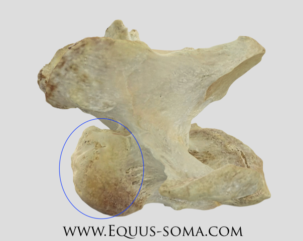
Jersey had navicular disease AND navicular syndrome (yes, there is a difference). He had two mobile (free-moving) chips off of the navicular bone along with a cyst. There was sclerosis around the chips which is indicative of some attempt at healing.
Fractures in the sixth rib on both sides. The fracture on the right sixth rib also had a sharp bony growth into the intercostal space of the fifth and sixth ribs.
When the team went to disarticulate some of the cervical spine, we discovered there was a malformation to the first rib in relation to where it should articulate with C7 (seventh cervical vertebra). It had an attachment to C7 and the head of the first rib had excess bony growth.
The sternum and T2 (second thoracic vertebra) showed evidence of dysplasia. His sternum wasn’t straight. He also had kissing spine through almost the entire late thoracic spine (I mistakenly did not write down the specific range of vertebrae- we can confirm this after composting)
5 lumbar vertebrae present, on their way to fusion in the dorsalis spinous processes aka DSPs (the bony projection at the top of the bone). His entire lumbar region was malformed with the DSPs being much larger than appropriate and all of the DSPs overriding one another but not yet fused. The sacroiliac junction could only be described as “nasty” due to its abnormality. It is possible that L5 & L6 were already completely fused. - we will know more here after he is fully composted.
The nerves in his lumbosacral area were “rearranged” and abnormal.
He had an abnormal posterior tilt of his pelvis which inverted when his head was brought down (unusual) and the right hip displayed arthritis. There was tissue degeneration that was indicative of an injury to the area.
Both hip joints showed significant cartilage degradation and synovitis (damage/degradation of synovial membrane).
His biceps femoris muscle appeared abnormal with a couple of areas of flaccid and fibrotic tissue. There was a scar tissue layer seemingly around the rupture in the tissue and the rupture in the tissue caused the surrounding muscle to be flaccid. The lateral patella ligament here appeared stretched with additional ruptures present.
Sesamoiditis developing in the left hind (bone chips starting to occur). Damage to the fetlock was also present- cartilage degradation throughout the limb. Additionally, the joint capsules were hyperemic (excess blood vessels) throughout.
The Cervical Spine:
Before the sixth (C6) and seventh (C7) cervical vertebrae were fully disarticulated, I did have a chance to watch the beginning of the disarticulation process. During this process, the team noted the C6 was awfully “short” (in terms of its width when looking at it from the side of the neck) for what it should have been.
Jersey had what is called “discospondylosis” (disc degeneration with erosion of the underlying bone) and osteoarthritis of the articular processes (APs). “It is highly likely that there was some impingement on the spinal cord. The growths on the articular processes (osteoarthritis) are formed in response to stresses and excessive, repetitive loading via the muscles that attach to them. Since there was extreme bone growth (on the C6 & C7) resulting in asymmetry between the two sides, this implies that Jersey’s body was trying to stabilize the region.” (From Ms. Ecklebarger).
Below are the notes (in italics) from Ms. Pamela Ecklebarger. She disarticulated these two vertebrae (along with Ms. Diane), processed them and made them into these incredible 3D models. Please read her notes here first and then you can click the link to view the models.
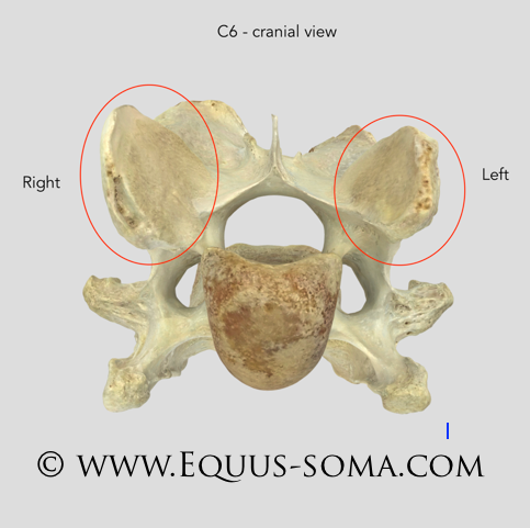
Cranial View of C6 (looking from the head towards the tail)
The red ovals outline the articular processes (APs). Note the obvious difference in size and shape. The right AP is much larger than “normal”. The bone on the edge of the left AP crumbled during processing. These form synovial joints with the APs of the neighboring vertebrae and should be of near or equal size and shape.
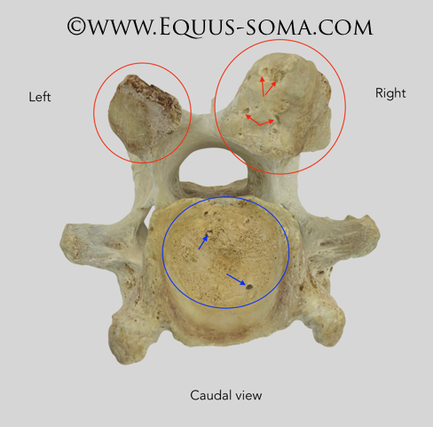
Caudal View of C6 (looking from tail toward the head)
You can really see the difference in sizes between right and left APs. Note also the “pits” (arrows) in the surfaces of these joints not only in the right AP but in the intervertebral joint as well (blue). The intervertebral joint is the main connection between the vertebrae by way of a disc. Sadly, in Jersey, the disc was degenerating or missing altogether between C6 and C7 = bone on bone. The normal surfaces of these joints should be smooth.
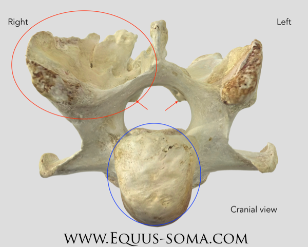
Cranial View of C7 (looking from the head towards the tail)
Extreme bony changes of the right AP. Red arrows point to bony growth that extends into the spinal cord space. More bony changes to the intervertebral joint (blue).
Lateral View of C7 (looking from the left side of the horse at the side of the cervical vertebra).
This lateral view illustrates the lumpy, bumpy (scientific term) surface of the cranial part of C7 that forms the intervertebral joint (blue oval). Again, this is supposed to be nice and smooth.
OK, now to the fun part… the 3D models. When you click or tap on the links below, a webpage will open with the 3D model in a window. Click/tap on the double-headed arrow in the lower right corner to open the model into full screen. Using the mouse or finger (tablet) you can zoom in/out, and grab the model to roll it all around. The screenshots above should help you orient and identify the structures.
This link to C6 opens in cranial view: https://sketchfab.com/3d-models/jersey-c6-b159b27165de4227bf87a84cfaddeb34.
This link to C7 also opens in cranial view: https://sketchfab.com/3d-models/jersey-c7-0d73fedb8b5e4cd0982291d72f00fe20.
Some Good (less scientific) Notes:
Jersey’s body was (for lack of a better term) a mess… Despite this, there was significant evidence that the things I was doing (nutrition, care, bodywork) were likely helping on a very MINIMAL scale but nevertheless, helping. His body was also doing an incredible job compensating– how magical is the body to just decide “I’m going to grow some bone here to fix an issue there?” or “I’m going to develop adipose tissue here to support the damage there” — SO COOL. Also, so incredibly important for bodyworkers to know when NOT to do work so we don’t interfere with beneficial compensations.
His body condition, as described by the team, was “perfect”. Despite the wacky ways his body was working to compensate, the team noted he had the correct amount of fat & muscle for his size.
There was healthy synovial fluid and healthy adipose tissue in some joints where there were OCD lesions. His body was trying to heal (important to note that this is not enough healing to outweigh the issues in his body- just nice to know my efforts were not wasted).
The opalescence of the bones, joints and fascia were all good signs. The health of his muscle tissue was great considering everything else that was happening internally.
He had a bolus in his esophagus. A bolus is what is formed as a horse finishes chewing so it can travel down into the stomach- Jersey was munching away (happily) on grass even in his last moments.
My Speculations: (These are my personal speculations. I am not a vet, I am not diagnosing. These speculations are based on observations of his body as he was dissected.)
The injuries to the ribs and sternum were indicative of birth canal trauma, or we can bundle them into the “rotational fall theory” noted below.
The evidence to support a rotational fall can be seen in the small healed fracture in the atlas (C1), the damage to the spleen, the scar tissue and damage in the front left shoulder, and the damage to the left hip.
In regards to the Cervical Spine— Some papers suggest that the disc degeneration is a result of infection or trauma. Because of these papers, it seems plausible that the trauma of a rotational fall could have been to blame for the issues (shortened C6, growths on the APs (articular processes), impingement on the spinal cord in the C6/C7 region causing neurological symptoms. Again- speculation.
Jersey’s vessel revealed what the experts noted as, “repetitive information”, when it came to his OCD lesions, wear and tear on his joints, stomach (old ulcers), lungs and heart. The injection site damage and damage to the liver from drug injections were not only news to me but an unfortunate reminder that the horse industry (not just racing) has a long way to go when it comes to managing drug use. This is NOT an accusation against whoever felt the need to pump drugs into him in his track days, but rather a reminder- to ALL of us, that LISTENING to our horses is the only way forward… the temporary “band-aid” type fixes— like unnecessary drugs to make a horse comfortable enough to compete through the pain— are NEVER okay.
In Conclusion:
I fully speculate that Jersey had a rotational fall in the early years of his life, likely before his track days, which affected some of the ongoings in his cervical spine and the issues in his left shoulder and hip. However, I also speculate that there were some congenital spinal issues because of the findings in his thoracic and lumbar regions- this I’ll never know for sure. How my horse walked, trotted, cantered, and GALLOPED soundly in his pasture and in his limited time under saddle will always blow my mind. The left front navicular bone had chipped off into multiple pieces which were freely existing as they pleased in his hoof capsule… and yet not a lame step. Jersey’s ability to remain so kind, loving, forgiving and warm will forever haunt me. Frankly, with everything going on in Jersey’s body, I am shocked he was not aggressive and didn’t try to kill me as I asked him to do work his body clearly couldn’t handle.
This is just the tip of my iceberg- between the experts, trainers, vets and friends that have helped me through this process, I feel I am just getting started. My heart and soul have been broken and slowly pieced back together… not just by these finding but as I work with others to help their horses find more comfort and helping their owners learn to listen. I will never stop loving this perfectly imperfect horse, for he has given me the greatest gifts in science, spirituality, and sparking a fire in me to do everything I can so that one less person or horse goes down the path Jersey and I did.
- With love and light, Katie